

Systemic Hypertension in Dogs and Cats: What's New?
Key ACVIM takeaways on blood pressure measurement and diagnosing systemic hypertension in dogs and cats, from measurement technique to treatment decisions.
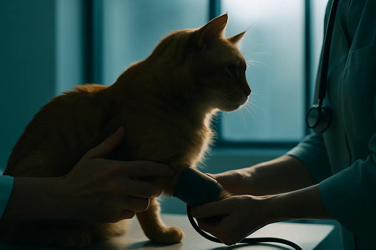
The most recent ACVIM meeting raised several points that challenge how blood pressure (BP) is measured and how systemic hypertension (SH) is diagnosed in dogs and cats. The gold standard for this measurement is central (direct) assessment, yet in everyday practice the devices used are Doppler and oscillometric units. To deliver reliable readings, these devices must be validated for each animal species, and the operator's experience also has a major influence on the accuracy of the results.
Setting and Measurement Technique
The setting chosen to collect the readings and the patient's emotional state have a strong influence on the results. Situational hypertension, triggered by anxiety or excitement, must be ruled out before confirming a diagnosis of SH. To minimize these interferences, measurements should be taken away from other patients, after the patient has acclimated to the room for at least 5 to 10 minutes and, whenever possible, with the owner present. The cuff must be matched to the animal's size, and each measurement session should include 5 or more repeated readings.
Causes and Diagnostic Workup
As noted, BP can rise in situations involving stressors, in association with underlying disease (secondary hypertension), or in the absence of any disease (idiopathic hypertension). In the idiopathic form, test results are typically normal, with no abnormalities in the complete blood count, serum biochemistry or urinalysis. The diagnostic workup may include abdominal ultrasound, serum symmetric dimethylarginine (SDMA), glomerular filtration rate (GFR), quantitative assessment of proteinuria, serum thyroxine concentration (cats only) and cortisol (dogs). Additional testing may include serum and urinary aldosterone as well as catecholamine concentrations.
Target-Organ Damage
Over time, SH leads to tissue injury and organ damage, which is what warrants treatment. The most commonly affected organs are the kidneys, where it progresses alongside chronic kidney disease; the eyes, with hyphema, hypertensive retinopathy and choroidopathy; the brain, where cerebrovascular events present as seizures, mental changes and unexplained neurological deficits; and the heart and blood vessels, resulting in left ventricular hypertrophy, left-sided congestive heart failure (uncommon) and aortic aneurysm.
Diagnosis and Risk Classification
A diagnosis of SH must be confirmed by a reliable BP measurement, and treatment decisions are based on those results. The presence of target-organ damage justifies starting treatment. In cases of prehypertension (140 to 159 mmHg) or moderate-risk SH (160 to 179 mmHg), measurements should be repeated every 4 to 8 weeks. In animals with severe SH (above 180 mmHg) the risk of organ damage is high, and measurements should be repeated weekly or, at most, every two weeks.
Treatment
Moderately or severely hypertensive animals should be treated, with the goal of bringing BP back to a prehypertensive or normotensive status. In dogs and cats, SH is frequently secondary (over 80% of cases); accordingly, antihypertensive therapy should be started together with treatment of the underlying cause. Beyond the underlying cause, the organs affected by SH must also be managed.
When treatment of the underlying cause is effective, the elevated BP may resolve completely or partially. The decision to use antihypertensive drugs should integrate all clinically available information, always prioritizing the patient's well-being and quality of life.