

B2B buying guide: how to choose a veterinary ECG machine (6 or 12 leads, resolution and reporting)
Before comparing price, compare what determines exam quality: how many leads your routine needs, which resolution and sampling rate preserve the signal, how the device handles artifact, and what happens to the tracing after capture, who interprets it and where the report is stored.
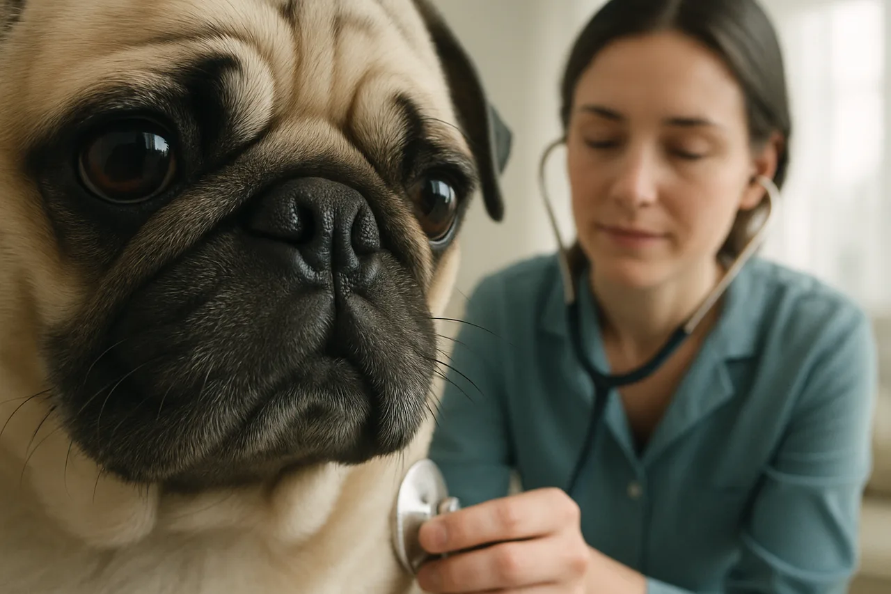
To choose a veterinary ECG machine, weigh six objective criteria before price: the number of leads your routine requires (6 frontal-plane leads resolve rhythm, rate and most arrhythmias; 12 leads add the precordials for spatial characterization, axis and conduction disturbances); resolution and sampling rate, which define how much of the real signal reaches the tracing; artifact and noise handling, since interpretation depends on a stable baseline; the capture format and battery autonomy across the clinic, the field and the surgical suite; who will interpret the tracing, whether there is a cardiologist in house or the report will come through telecardiology; and record integration, that is, what happens to the exam after capture. No device replaces interpretation: diagnosis and management always rest with the veterinarian.
1. How many leads your routine requires
The question is not which device has the most leads, but how many your routine actually uses. The 6 frontal-plane leads (I, II, III, aVR, aVL and aVF) suffice to determine rate, rhythm, mean electrical axis and to recognize most arrhythmias, which is what screening, pre-anesthetic assessment and follow-up of cardiac patients require day to day.
Twelve leads add the precordials, which provide the spatial reading of the cardiac vector. They help in finer characterization of chamber overload, in locating ectopic foci and in conduction disturbances, the typical setting of a complete cardiology workup and of referred cases. If the practice screens and refers complex cases to a cardiologist, 6 leads usually suffice; if the practice itself runs the workup, 12 leads pay for themselves.
2. Resolution and sampling rate: what preserves the signal
Resolution and sampling rate define how much of the real electrical signal survives into the tracing you read. In dogs and cats this matters more than in humans: heart rates are higher, complexes are narrower and deflections lasting a few milliseconds carry diagnostic information. A low sampling rate smooths away precisely those fine details, and low resolution loses amplitude in low-voltage tracings.
The practical benchmark comes from the normal values themselves: in dogs, QRS duration in lead II runs to about 0.06 second in small and medium breeds and up to 0.07 second in large and giant breeds, and the P wave lasts about 0.04 second (Santilli et al.). Measuring a complex of that order reliably requires a generous sampling rate. Ask the manufacturer for the explicit figures of resolution in bits and samples per second per channel, and compare them across candidates.
3. Artifact, noise and standardizing technique
A tracing is only interpretable if the baseline is stable. Muscle tremor, patient movement, poor electrode contact and mains interference are the classic sources of artifact, and no filter fully corrects a signal captured under poor conditions. It is therefore worth assessing how the device flags poor contact, which filters it offers and how tolerant capture is with an awake patient.
There is a useful experimental data point for this decision: a study comparing recording devices, body position, electrode placement and sedation showed that these variables affect electrocardiogram intervals in dogs (Bogucki and Noszczyk-Nowak; see also Rishniw et al.). The practical reading is that standardizing technique weighs as much as device specification, and that switching equipment or protocol during follow-up of the same patient calls for caution when comparing intervals.
4. Who reads the tracing: in-house analysis or telecardiology
Capturing is half the exam; the other half is interpreting and recording. Here the decisive question is who reads the tracing. If there is a cardiologist on staff, the weight falls on the analysis tools: lead-by-lead measurement, axis calculation, continuous recording to review the whole exam and report generation. If there is not, the criterion becomes how the exam reaches a specialist.
Telecardiology is already described in the veterinary literature as viable for remote interpretation: in home monitoring with ECG sent for remote analysis, most received tracings were deemed interpretable and inter-observer agreement on rhythm diagnosis was good (Vezzosi et al.). The message for purchasing is twofold: remote reporting works, but it depends on the quality of the captured signal, which reinforces the earlier criteria. In Brazil, such a report is telediagnosis regulated by CFMV Resolution No. 1.465/2022 and requires a veterinarian with active registration in the CFMV/CRMV system; equivalent rules apply in other jurisdictions.
5. Record integration: what happens after capture
The last criterion is the one usually forgotten in the comparison and the one that hurts most later: where the exam ends up. A tracing locked in a local file on the front-desk computer does not support longitudinal follow-up, does not travel with the patient on referral and does not allow comparing today exam with the one from six months ago. Assess whether the exam uploads linked to the patient record, whether the history is reachable from another device, and whether sending for a remote report is part of the same flow or an improvised email attachment.
| Criterion | Screening and routine practice | Complete cardiology workup |
|---|---|---|
| Leads | 6 (frontal plane) resolve rhythm, rate and most arrhythmias | 12 simultaneous add the precordials (axis, conduction, chamber overload) |
| Resolution and sampling | ask for the figure in bits and in samples per second per channel | the higher, the more faithful to deflections of a few milliseconds |
| Interpretation | telecardiology when there is no cardiologist on staff | in-house tools: measurement, axis, continuous recording, reporting |
| Where the exam ends up | linked to the patient record, with reachable history | same flow as sending for a remote report |
How to decide in practice
Bringing the six criteria together: first define whether your routine calls for 6 or 12 leads, demand resolution and sampling figures from the manufacturer instead of adjectives, test capture on the awake patient you actually see, standardize positioning technique, decide who will interpret and verify that the exam ends up linked to the patient record. Price comes after these filters, not before them. If the question is whether to buy the device or work with a lease that includes remote reporting, we cover that comparison in a dedicated guide.
Sources
- Santilli R, Moïse NS, Pariaut R, Perego M. Electrocardiography of the Dog and Cat: Diagnosis of Arrhythmias. 2nd ed. Edra; 2018. (2018)
- Effects of recording device, body position, electrode placement, and sedation on electrocardiogram intervals in dogs (2022) PMID 36028186
- Home monitoring of heart rate and heart rhythm with a smartphone-based ECG in dogs (2018) PMID 30559174
- Rishniw M, Porciello F, Erb HN, Fruganti G. Effect of body position on the 6-lead ECG of dogs. J Vet Intern Med. (2002) PMID 11822807
- Resolução CFMV nº 1.465, de 27 de junho de 2022 (regulamenta a telemedicina veterinária, incluindo telediagnóstico) (2022)