

Pulse Oximetry (SpO2) in Dogs and Cats: What It Measures, What It Misses, and How to Read the Waveform
Pulse oximetry continuously and non-invasively estimates hemoglobin oxygen saturation (SpO2), but it does not replace capnography or assess ventilation. Understand reference ranges, the dissociation curve, reading the plethysmographic waveform, and practical limitations in anesthesia, emergency, and hospitalization.
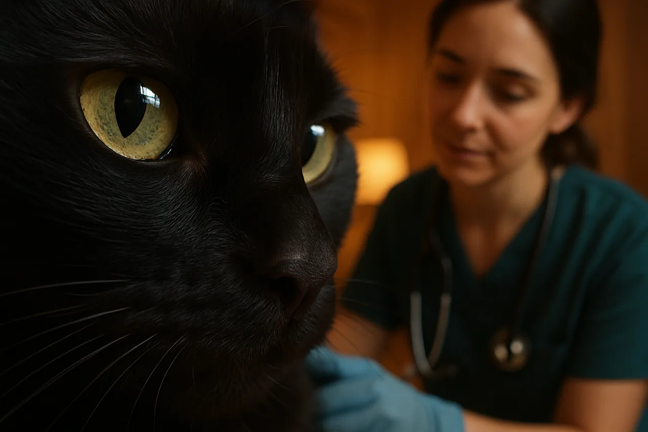
Pulse oximetry is a continuous, non-invasive method that estimates the oxygen saturation of arterial hemoglobin, expressed as SpO2 in percent, in dogs and cats. It tells you how much of the circulating hemoglobin is carrying oxygen, but it does not directly measure the partial pressure of oxygen (PaO2) in blood or ventilation, meaning it does not indicate whether the patient is clearing carbon dioxide adequately. For this reason, pulse oximetry complements but does not replace capnography and arterial blood gas analysis. In the ACVAA small animal anesthesia monitoring guidelines (2025), when supplemental oxygen is being delivered, SpO2 is considered a late indicator of an oxygenation problem, and any value below 95% should be investigated, both to assess the patient and to rule out technical issues. The final clinical decision always rests with the attending veterinarian, integrating the number, the waveform, and the animal's overall picture.
What pulse oximetry actually measures
The sensor emits light at two wavelengths (red and infrared) through a pulsatile tissue bed. Oxyhemoglobin and deoxyhemoglobin absorb these wavelengths differently, and the device calculates the ratio between them during arterial pulsation, producing the SpO2. The pulsatile component is precisely what allows arterial blood to be isolated from venous blood and static tissue.
It is important to separate two concepts: SpO2 (the optical estimate from the pulse oximeter) and SaO2 (the true saturation measured by co-oximetry on a blood gas sample). Laboratory co-oximetry, validated in healthy dogs, distinguishes several hemoglobin species, including oxyhemoglobin, deoxyhemoglobin, carboxyhemoglobin, and methemoglobin. A conventional two-wavelength pulse oximeter does not make this distinction, which is why it can overestimate saturation in the presence of carboxyhemoglobin (carbon monoxide poisoning) or behave anomalously with methemoglobin.
Reference ranges and the hemoglobin dissociation curve
In dogs and cats breathing room air and well oxygenated, the expected SpO2 sits around 95% to 100%. Values below 95% warrant investigation, and broadly speaking an SpO2 around 90% already corresponds to a PaO2 near 60 mmHg, the classic threshold of hypoxemia that demands immediate attention. These numbers must be interpreted by the veterinarian in context: supplemental oxygen, altitude, and hemodynamic status all change the reading.
The relationship between SpO2 and PaO2 is not linear: it follows the S-shaped oxyhemoglobin dissociation curve. On the upper plateau of the curve (high saturations), large drops in PaO2 produce only small changes in SpO2, which explains why oximetry is a late indicator when the patient is receiving oxygen. On the steep portion of the curve (below roughly 90%), small drops in PaO2 cause saturation to fall rapidly. Factors such as temperature, pH, carbon dioxide, and 2,3-DPG shift the curve, altering hemoglobin's affinity for oxygen at the same SpO2.
Reading the plethysmographic waveform: the same logic as reading the signal
Just as important as the number is the plethysmographic waveform that accompanies it. This curve represents the arterial pulsation detected by the sensor and acts as the primary indicator of signal quality: a regular waveform with consistent amplitude and morphology, synchronized with the patient's actual pulse, supports confidence in the displayed SpO2. As with an ECG tracing, the clinician should trust the waveform before trusting the value.
When the plethysmographic waveform is flattened, irregular, noisy, or mismatched with the auscultated or palpated pulse rate, the SpO2 value should be treated with caution and likely reflects an artifact rather than physiology. Checking whether the oximeter's pulse rate matches the animal's heart is a simple and powerful safeguard against false readings. For this reason, multiparameter monitors display the waveform and a variable-pitch audible pulse tone, recommended by the ACVAA guidelines so the team can detect saturation drops even without looking at the screen.
Practical limitations and how to work around them
Pulse oximetry depends on an adequate pulsatile signal, and several situations common in practice degrade that reading. Hypoperfusion, hypothermia, and vasoconstriction reduce pulsatile flow and can zero out or make the value erratic; movement and shivering introduce artifact; heavy pigmentation, dark mucous membranes, and thick hair coats interfere with light transmission; and strong ambient light (surgical lamps, phototherapy) can contaminate the sensor. Carboxyhemoglobin and methemoglobin, in turn, distort the saturation estimate of conventional oximeters.
Strategies to work around these limitations are practical: try different sensor sites (tongue, lip, skin fold, ear, toe, base of the tail, or metatarsus), switch between transmission and reflectance sensors, keep the patient warm and well perfused, reduce movement, shield the sensor from ambient light, and rotate the site to avoid pressure injury during prolonged use. Studies in anesthetized dogs show that different sites and sensor technologies can provide agreeing SpO2 readings, but they also reinforce that reliability depends on adequate perfusion and positioning. Whenever there is any doubt about oxygenation or ventilation, the correct approach is to confirm with arterial blood gas analysis and capnography.
Use in anesthesia, emergency, and hospitalization
In anesthesia, pulse oximetry is part of the minimum oxygenation monitoring and is associated with a reduced risk of perianesthetic mortality. Because, under supplemental oxygen, it detects desaturation late, it should be used alongside capnography, observation of mucous membrane color, and hemodynamic monitoring, with audible alarms set. An SpO2 below 95% during anesthesia requires immediately checking both the patient and the equipment.
In emergency and hospitalization, SpO2 helps triage and follow patients with respiratory disease, shock, or postoperative status, offering a continuous oxygenation trend without drawing blood. The reading, however, must be interpreted cautiously in hypoperfused, agitated, or hypothermic animals, precisely the most common in emergencies, where the signal can fail. The isolated value never replaces the clinical exam or blood gas analysis when hypoxemia or a ventilation disturbance is suspected; it guides, but the decision always belongs to the veterinarian.
Frequently asked questions
Does pulse oximetry replace capnography? No. SpO2 assesses hemoglobin oxygenation, while capnography assesses ventilation and carbon dioxide clearance; these are distinct, complementary pieces of information. A patient can maintain a high SpO2 on oxygen and still be ventilating poorly, with CO2 retention that only capnography would reveal.
Does an SpO2 of 100% guarantee everything is fine? Not necessarily. Under supplemental oxygen, saturation can remain high even as oxygenation worsens, because of the plateau of the dissociation curve; this is why oximetry is considered a late indicator. One should always assess the plethysmographic waveform, the clinical picture, and, when indicated, blood gas analysis.
Why does the oximeter show values that do not make sense? The most common causes are technical: poor perfusion, movement, vasoconstriction, inadequate sensor positioning or site, heavy hair coat or pigmentation, and ambient light. Check that the waveform is clean and that the device's pulse rate matches the animal's heart before trusting the number.
Sources
- The American College of Veterinary Anesthesia and Analgesia Small Animal Anesthesia and Sedation Monitoring Guidelines 2025 (2025) PMID 40447502
- Co-oximetry in clinically healthy dogs and effects of time of post sampling on measurements (2011) PMID 21981712
- Comparison of human smartwatch and transmittance pulse oximetry for evaluating peripheral oxygen saturation in anesthetized dogs (2024) PMID 39624191