

ECG in Cats and Feline Hypertrophic Cardiomyopathy (HCM): Why the Cat Hides the Disease
Cats are masters at hiding heart disease. Understand the real role of the ECG in feline hypertrophic cardiomyopathy (arrhythmias and clues of chamber enlargement), why echocardiography is the definitive test, and what to weigh in pre-anesthetic screening of the asymptomatic cat.
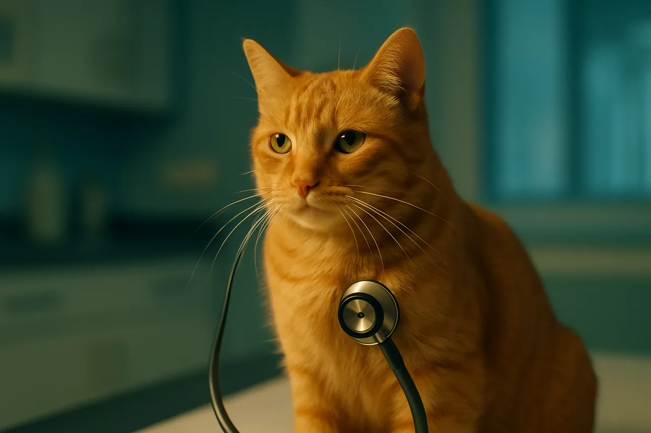
Feline hypertrophic cardiomyopathy (HCM) is the most common acquired heart disease in cats, defined by thickening of the left ventricular muscle with no identifiable cause, and it is estimated to affect roughly 15% of the general cat population. The electrocardiogram (ECG) records the heart's rhythm and electrical activity: it helps identify arrhythmias and raises clues of chamber enlargement, but it does not diagnose HCM. The definitive test is echocardiography, which directly measures myocardial thickness.
Why the cat hides heart disease
The cat is a silent patient. Most felines with HCM remain asymptomatic for months or years, with no cough and few signs noticeable to the owner, and many cases only surface when the veterinarian hears a murmur or a gallop sound during a routine visit. The catch is that auscultation has low sensitivity: it is estimated that at least a third of cats with cardiomyopathy, and about half in some series, have no murmur at all, and a share of cats with a soft murmur have no structural heart change.
This behavior hides a real risk. The first manifestation of HCM can be dramatic and sudden: congestive heart failure (CHF) with breathing difficulty, arterial thromboembolism (ATE, the clot that often paralyzes the hind limbs, with intense pain), or sudden death. Relying on symptoms alone to suspect feline heart disease is therefore a fragile strategy: by the time clinical signs appear, the disease is usually already advanced.
The role of the ECG: what it shows (and what it does not)
The ECG is the tool of choice for characterizing cardiac rhythm. In cats with HCM, premature ventricular complexes, ventricular tachycardia, premature supraventricular complexes, second- and third-degree atrioventricular block, and sinus bradycardia may arise. The tracing can also suggest chamber enlargement: tall R waves in lead II are associated with left ventricular hypertrophy, and tall or widened P waves suggest atrial enlargement. Documenting an arrhythmia is clinically meaningful, and the reverse correlation is good: in one series, 102 of 106 cats with ventricular arrhythmias on ECG had echocardiographic changes consistent with cardiomyopathy.
The major limitation is the ECG's low sensitivity for HCM. In one study, the presence of arrhythmias on a 2-minute recording had a sensitivity of only 31% and a specificity of 100% for identifying left ventricular hypertrophy, and about 43% of cats with HCM had an ECG with no relevant abnormalities. In other words: a normal ECG does not rule out HCM. The ECG is therefore more useful for confirming and classifying suspected arrhythmias (and, when indicated, 24-hour Holter monitoring extends that capture) than for screening for the structural disease itself. The decision on which tests to order and how to interpret them always rests with the attending veterinarian.
Echocardiography: the definitive diagnosis of HCM
If the ECG watches the rhythm, echocardiography watches the structure. The ACVIM consensus (2020) defines echocardiography as the reference test for HCM: it measures left ventricular wall thickness, identifies the pattern of hypertrophy, assesses left atrial size (an important risk marker), and detects outflow tract obstruction. The ECG and the echocardiogram do not compete, they complement each other: the first characterizes the rhythm, the second confirms and stages the disease.
The ACVIM also proposes a staging system that separates cats with subclinical cardiomyopathy into lower and higher risk of complications, which guides monitoring and management. Biomarkers such as NT-proBNP can support screening by indicating which cats warrant an echocardiogram, but they do not replace imaging. Diagnosis, staging, and the treatment plan are defined by the veterinarian, ideally with the support of a cardiologist when the case requires it.
Pre-anesthetic screening of the cat
Occult HCM is one of the biggest concerns before feline anesthesia. An apparently healthy cat, scheduled for a neuter or a dental cleaning, may have subclinical heart disease that alters tolerance to fluid therapy and anesthetic agents. The AAFP feline anesthesia guidelines (2018) reinforce the importance of careful pre-anesthetic evaluation, including attentive auscultation for murmur and gallop.
When findings such as a murmur, gallop sound, or arrhythmia are present, deepening the workup before an elective procedure is advised, with complementary tests such as echocardiography and, when appropriate, NT-proBNP, according to clinical judgment. Here the ECG has practical value: monitoring rhythm in the peri- and intra-anesthetic period allows early recognition of arrhythmias and instability. The depth of this screening (and the decision to postpone or proceed with anesthesia) depends on the patient, the procedure's risk, and the veterinarian's judgment.
The asymptomatic cat hook: watch before the event
The numbers reinforce why the asymptomatic cat deserves attention. In the international REVEAL study, with more than a thousand cats with preclinical HCM, the 1-year risk of CHF and ATE was about 7.0% and 3.5%, rising to roughly 19.9% and 9.7% at 5 years, with cardiovascular mortality around 22.8% at 5 years. These are cats with no symptoms at the start of follow-up that nonetheless carry substantial risk over time.
The practical message is one of vigilance, not alarm: identifying a murmur or gallop at the visit, investigating with the appropriate tests, and following the at-risk cat allows action before the acute event. The ECG comes in as one piece of the picture, characterizing the rhythm and supporting monitoring; echocardiography closes the diagnosis. No single test replaces the full evaluation, and every decision rests with the veterinarian responsible for the patient.
Sources
- Fuentes VL, Abbott J, Chetboul V, et al. ACVIM consensus statement guidelines for the classification, diagnosis, and management of cardiomyopathies in cats. J Vet Intern Med. 2020;34(3):1062-1077. (2020) PMID 32243654
- Cofaru A, Murariu R, Popa T, Peștean CP, Scurtu IC. The Unseen Side of Feline Hypertrophic Cardiomyopathy: Diagnostic and Prognostic Utility of Electrocardiography and Holter Monitoring (review). Animals (Basel). 2024;14(15):2165. (2024) PMID 39123690
- Fox PR, Keene BW, Lamb K, et al. International collaborative study to assess cardiovascular risk and evaluate long-term health in cats with preclinical hypertrophic cardiomyopathy and apparently healthy cats: The REVEAL Study. J Vet Intern Med. 2018;32(3):930-943. (2018) PMID 29660848
- Robertson SA, Gogolski SM, Pascoe P, Shafford HL, Sager J, Griffenhagen GM. AAFP Feline Anesthesia Guidelines. J Feline Med Surg. 2018;20(7):602-634. (2018) PMID 29989502
- Cornell Feline Health Center. Hypertrophic Cardiomyopathy (HCM). Cornell University College of Veterinary Medicine.
- Prevalence and Clinical Significance of Heart Murmurs Detected on Cardiac Auscultation in 856 Cats (Ferasin L, et al.). Veterinary Sciences. 2022. (2022) PMID 36288177