

Main arrhythmias in dogs and cats: how to recognize them on the ECG
From respiratory sinus arrhythmia (normal in dogs) to atrial fibrillation, atrioventricular blocks and VPCs: a source-anchored clinical guide on how to spot each arrhythmia on the ECG and when a case is an emergency or warrants referral to a cardiologist.
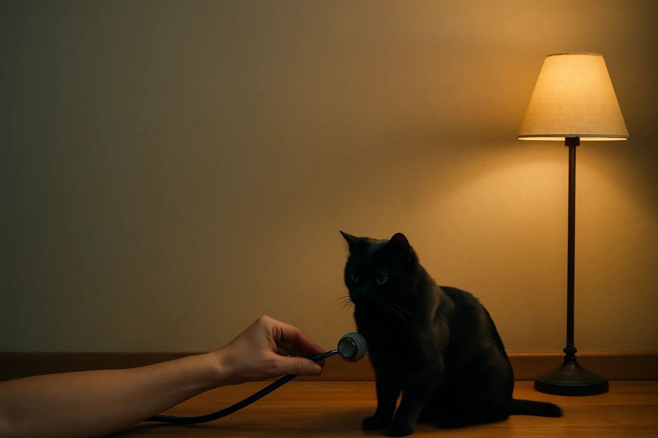
An arrhythmia is any change in the heart's rhythm, rate or electrical conduction that departs from normal sinus rhythm. In dogs and cats, the most frequent in clinical practice are respiratory sinus arrhythmia (a normal variant in dogs), atrial fibrillation, atrioventricular blocks (1st, 2nd and 3rd degree) and ventricular premature complexes (VPCs). The electrocardiogram (ECG) is the test that confirms the type of arrhythmia, because different rhythms can sound alike on auscultation. This article is educational in nature: tracing interpretation and any clinical decision rest with the veterinarian responsible for the case.
First things first: what normal rhythm looks like
In normal sinus rhythm, every beat arises in the sinus node, there is one P wave for each QRS and one QRS for each P wave, and the P-R interval is constant. Reference heart-rate ranges are commonly cited around 60 to 170 bpm in dogs and 140 to 220 bpm in cats, bearing in mind that a stressed cat in the exam room can reach 240 bpm. These values vary with body size, age and stress level, and must be interpreted in the context of each patient.
Respiratory sinus arrhythmia: the normal finding that alarms
Respiratory sinus arrhythmia is a physiological variation of sinus rhythm: heart rate rises on inspiration and falls on expiration, reflecting high vagal tone. In dogs it is a normal and even expected finding; in the hospitalized cat it is usually uncommon and may suggest underlying disease, although it appears more often in the home environment.
On the tracing there is a regularly irregular rhythm, with R-R interval variation typically above 10%, keeping one P wave for each QRS. P-wave morphology may shift slightly (wandering pacemaker). A practical point: sinus arrhythmia is abolished by an increase in rate (excitement, exercise) or by vagolytic drugs such as atropine, which helps distinguish it from conduction blocks.
Is it an emergency? No. It is a benign finding. The caution is not to mistake the pauses for atrioventricular block; when the nature of a pause is uncertain, or in a cat with marked sinus arrhythmia, it is worth recording a tracing and discussing the case.
Atrial fibrillation: an irregularly irregular, fast rhythm
Atrial fibrillation is characterized on the ECG by the absence of P waves, an undulating (fibrillatory) baseline and an irregularly irregular supraventricular rhythm with normal-looking QRS complexes. The ventricular rate is usually fast and irregular, with a cited range of 80 to 300 bpm in dogs; rates above 160 bpm generally signal a need for control. On auscultation, the rhythm has been likened to bongo drums or shoes tumbling in a dryer.
In most dogs and cats, atrial fibrillation is associated with structural heart disease and marked left atrial enlargement. In giant-breed dogs, so-called lone atrial fibrillation can occur without evident structural disease. The classic therapeutic goal is ventricular rate control, with targets often cited around 140 to 160 bpm and, on Holter, a mean rate below 125 bpm.
When is it an emergency or a referral? A very high, sustained ventricular rate can decompensate the patient and is potentially serious. Every confirmed atrial fibrillation warrants cardiology assessment with echocardiography to investigate the underlying disease and define a rate-control strategy, ideally with a cardiologist.
Atrioventricular blocks: when the impulse does not get through
Atrioventricular (AV) blocks reflect delay or failure of impulse conduction between atria and ventricles, and are classified by degree. In first degree, every impulse gets through but is delayed: the P-R interval appears prolonged and each P still conducts a QRS. It is often vagal and, in isolation, usually carries little clinical consequence.
In second degree, some atrial impulses are not conducted: there are P waves without a corresponding QRS. In Mobitz type I (Wenckebach), the P-R interval lengthens progressively until a blocked P; in Mobitz type II, the P-R interval stays constant before the blocked P. Mobitz I is usually vagal in origin and benign; Mobitz II and high-grade forms tend to be pathologic.
In third degree (complete AV block), no atrial impulse reaches the ventricles: there is complete dissociation between P waves and QRS, with the ventricles maintained by a slow escape rhythm. In dogs this escape is usually around 20 to 60 bpm; in cats it is cited at 80 to 130 bpm, which is why many cats show few signs.
When is it an emergency or a referral? Third-degree AV block and symptomatic high-grade blocks (syncope, weakness, exercise intolerance) are emergencies and call for urgent cardiology assessment; the definitive treatment in dogs is usually pacemaker implantation. An atropine test (cited at 0.04 mg/kg) helps distinguish a vagal cause from conduction disease, but the decision rests with the veterinarian/cardiologist.
Ventricular premature complexes (VPCs) and ventricular tachycardia
A ventricular premature complex (VPC) is a beat that arises in the ventricle, outside the sinus node, and comes early in the rhythm. On the ECG it appears as a wide, bizarre, premature QRS without an associated P wave in front of it. VPCs can occur singly, in pairs (couplets), in triplets or in longer runs.
When three or more ventricular premature complexes occur in sequence, it is called ventricular tachycardia. Features regarded as malignant include high rates (cited above 180 bpm), polymorphic morphology and the R-on-T phenomenon, in which the VPC falls on the T wave of the previous beat and can trigger ventricular fibrillation and sudden death, especially in hearts with myocardial disease (for example, Boxer and Doberman cardiomyopathies).
When is it an emergency or a referral? Isolated VPCs in an asymptomatic patient do not always require treatment, but the cause should be investigated (cardiac, electrolyte or systemic). In contrast, sustained ventricular tachycardia, very high rates, a polymorphic pattern, R-on-T or a symptomatic patient (weakness, syncope) constitute an emergency: acute control is usually achieved with intravenous lidocaine, and the case warrants referral to a cardiologist. Every therapeutic decision rests with the veterinarian.
Red flags: when it is an emergency and when to refer
Signs that raise suspicion of a significant arrhythmia and call for immediate assessment: syncope or collapse, marked weakness, exercise intolerance, pale or cyanotic mucous membranes, respiratory effort and a pulse that is very fast, very slow or clearly irregular. In the face of any of these, recording an ECG and stabilizing the patient are clinical priorities.
As a practical rule, higher-risk scenarios include: atrial fibrillation with a high, sustained ventricular rate; third-degree or symptomatic high-grade AV block; and sustained ventricular tachycardia or one with malignant features (very high rate, polymorphism, R-on-T). These scenarios, and any arrhythmia in a symptomatic patient or one with structural heart disease, justify referral to a cardiologist. The approaches described here are references from the literature, not a prescription: the final decision always rests with the responsible veterinarian.
The role of connected ECG
Telling these arrhythmias apart depends on a good-quality tracing and, often, on a specialist second opinion. INpulse ECG devices such as the INcardio X (12 leads) and the INcardio Agile (6 leads) capture the signal and integrate it with INcloud, allowing the exam to be archived, compared with previous tracings and sent for a cardiologist tele-report when the case requires. The technology supports clinical decision-making; it does not replace the veterinarian's judgment.
Sources
- Heart Disease: Conduction Abnormalities in Dogs and Cats (Merck Veterinary Manual)
- Top 5 Arrhythmias in Dogs & Cats, Treatment Protocols (Clinician's Brief)
- Interpreting ECGs With Confidence in Veterinary Medicine: Part 1 (Clinician's Brief)
- Third-Degree Atrioventricular Block - an overview (ScienceDirect Topics, Veterinary Science)
- ACVIM consensus statement guidelines for the classification, diagnosis, and management of cardiomyopathies in cats (2020) PMID 32243654
- ACVIM consensus guidelines for the diagnosis and treatment of myxomatous mitral valve disease in dogs (2019) PMID 30974015
- Vagally Associated Second Degree Atrio-Ventricular Block in a Dog with Severe Azotemia and Evidence of Sympathetic Overdrive (2022) PMID 35622751
- A study on the electrocardiography in dogs: Reference values and their comparison among breeds, sex, and age groups (2020) PMID 33281359