

Veterinary Capnography: The Complete Guide for Clinical Practice
Veterinary capnography measures exhaled CO₂ (EtCO₂) to assess a patient's ventilation. Learn the core concepts, mainstream vs. sidestream modules, its role in anesthesia and how to read the waveform.
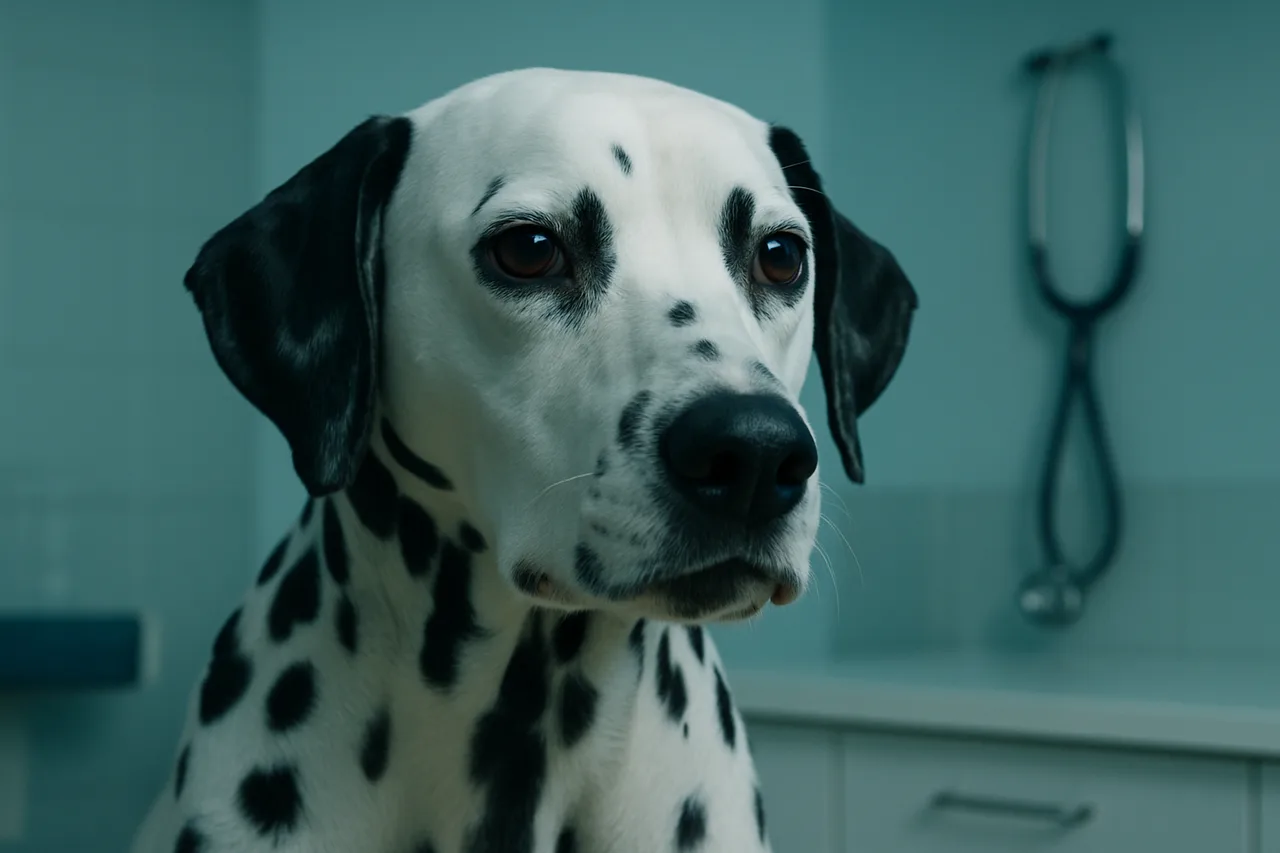
Veterinary capnography is an essential tool in patient monitoring, widely used across clinical scenarios to assess a patient's respiratory efficiency. This non-invasive method delivers valuable insight into gas exchange and metabolic status, supporting decision-making during anesthetic procedures, emergencies and critical care.
In this article we walk through the core concepts, benefits and applications of veterinary capnography, along with practical guidance on interpreting results and choosing the right equipment.
What is capnography and how does it work?
Capnography is the measurement of the partial pressure of carbon dioxide (CO₂) at the end of exhalation, known as EtCO₂. It directly reflects the patient's alveolar ventilation and its ability to eliminate CO₂. The measurement is displayed as a waveform or numeric values, letting the veterinarian continuously monitor the animal's respiratory status.
Capnography relies on sensors that detect the CO₂ present in exhaled air. There are two main types of capnographs, sidestream and mainstream, each with specific applications, as we will see below.
What are the differences between mainstream and sidestream modules?
1. Mainstream:
Measures CO₂ directly within the breathing circuit, with no need to collect air samples.
Delivers fast, accurate readings, making it ideal for patients that require continuous, real-time monitoring, such as large-breed animals.
It is more robust, but it can add weight to the breathing circuit, which calls for care in smaller patients.
2. Sidestream:
Draws a small sample of exhaled air through a sampling line connected to the breathing circuit.
It is lighter and more compact, well suited to practices monitoring multiple patients and to situations where a lightweight setup is essential.
It may show a slight delay in the reading compared with the mainstream module, but it offers greater flexibility in use.
What is the difference between capnography and oximetry?
Although capnography and oximetry are both used to monitor patients, they measure different parameters.
While capnography assesses exhaled CO₂, oximetry measures the oxygen saturation in the blood. Oximetry provides information about oxygenation, but it does not directly reflect the quality of ventilation.
These technologies are complementary in anesthetic monitoring and critical care. Combining the two gives a more complete picture of the patient's respiratory status, increasing safety during procedures and treatments.
Why does capnography matter in anesthetic monitoring?
Capnographic monitoring is indispensable during anesthesia because it provides crucial data on the patient's ventilation and perfusion. This is especially relevant in situations where the respiratory response may be altered by anesthetic drugs.
Benefits of using capnography in anesthesia:
Early detection of respiratory problems: capnography makes it possible to identify hypoventilation, apnea and airway obstruction before they become critical.
Identification of anesthetic complications: analyzing EtCO₂ helps recognize hemodynamic and metabolic changes that may signal complications.
Need for rapid intervention: capnography delivers real-time information, enabling immediate adjustments in emergencies.
Integrating capnography with other monitoring methods, such as oximetry and blood pressure, significantly increases the safety and quality of anesthetic care.
How to choose capnography monitoring equipment
Choosing the right capnograph depends on your clinical needs and the type of patient you treat. Here are the main types of equipment and their characteristics:
Sidestream capnograph: this type draws a small sample of exhaled air through a sampling line connected to the breathing circuit. It is light and compact, making it suitable for practices that monitor multiple patients.
Mainstream capnograph: this model measures CO₂ directly within the breathing circuit, with no need for additional sampling. It is ideal for large-breed patients or those that require more precise monitoring.
Multiparameter monitor: multiparameter devices combine capnography with other monitoring functions, such as oximetry, electrocardiogram and blood pressure. They are highly versatile and recommended for practices that want to centralize monitoring.
When making your choice, it is important to weigh factors such as value for money, equipment maintenance and ease of use.
How to interpret capnography results and parameters
Interpreting capnography requires understanding the parameters being measured and how they vary. The key value analyzed is EtCO₂, which typically ranges between 35 and 45 mmHg in healthy animals.
Changes in these values may indicate:
Elevated EtCO₂: may suggest hypoventilation, increased CO₂ production or airway obstruction.
Reduced EtCO₂: usually points to hyperventilation, a drop in cardiac output or inadequate perfusion.
The shape of the capnography waveform is also essential for interpretation. A normal waveform has three phases: 1. Baseline (inspiration with no CO₂); 2. Expiratory upstroke (initial CO₂ release); 3. Plateau (EtCO₂ stabilized at the end of exhalation).
Any change in the shape of the waveform can signal complications, such as a circuit leak, obstructions or a mechanical ventilator failure.
Conclusión
Veterinary capnography is an indispensable tool for respiratory monitoring across a wide range of clinical scenarios, ensuring greater safety and quality in patient care. With its ability to detect respiratory problems and anesthetic complications early, this technology stands out as an essential ally for veterinary clinics and hospitals.
When choosing capnography equipment, it is important to consider the specific needs of your clinical practice, ensuring a choice that delivers reliability and convenience. Beyond that, understanding and correctly interpreting the measured parameters enables fast, confident interventions that promote a better quality of life for patients.
Sources
- The American College of Veterinary Anesthesia and Analgesia Small Animal Anesthesia and Sedation Monitoring Guidelines 2025. (2025) PMID 40447502
- 2024 RECOVER Guidelines: Monitoring. Evidence and knowledge gap analysis with treatment recommendations for small animal CPR. (2024) PMID 38924672
- Quality Improvement: Diagnostic Accuracy of Mainstream Versus Sidestream Capnography in Detecting Airway Intubation of Small-Bore Styleted Nasoenteric Feeding Tubes in Dogs and Cats. (2025) PMID 40625308
- Agreement between values for arterial and end-tidal partial pressures of carbon dioxide in spontaneously breathing, critically ill dogs. (2009) PMID 19951100
- Accuracy of noninvasive oxyhemoglobin saturation, end-tidal carbon dioxide concentration, and blood pressure monitoring during experimentally induced hypoxemia, hypotension, or hypertension in anesthetized dogs. (1998) PMID 9492938